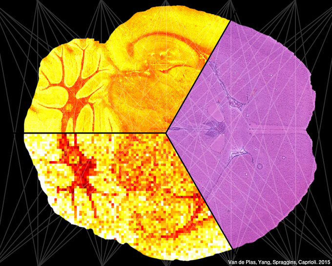
The Molecular Image Fusion system is a software framework that can predict ion distributions in tissue. It accomplishes this by fusing data from two distinct technologies: imaging mass spectrometry (IMS) and microscopy. IMS-generated molecular maps, rich in chemical information but having coarse spatial resolution, are combined with optical microscopy maps, which have relatively low chemical specificity but high spatial information. The resulting images combine the advantages of both technologies, enabling prediction of a molecular distribution both at high spatial resolution and with high chemical specificity. We have demonstrated the potential of image fusion through several applications: (i) ‘sharpening’ of IMS images, which uses microscopy measurements to predict ion distributions at a spatial resolution that exceeds that of measured ion images by ten times or more; (ii) prediction of ion distributions in tissue areas that were not measured by IMS; and (iii) enrichment of biological signals and attenuation of instrumental artifacts, revealing insights that are not easily extracted from either microscopy or IMS individually.
The predictive applications of image fusion can contribute in those cases where conducting a physical measurement is unpractical (e.g. due to measurement time, varying instrument stability), uneconomical (e.g. laser wear, detector deterioration), or even infeasible due to instrumental limitations (e.g. spatial resolution beyond laser capabilities, low SNR). The prediction methods that the Molecular Image Fusion system provides, and the cross-technology pattern discovery that drives them, can fulfill an instrumental role in realizing the full potential of multi-modal imaging studies, particularly in the molecular mapping of tissue.


